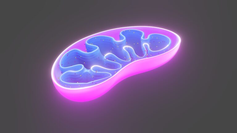
Sperm contains multiple mitochondria in the middle piece and in the tail that supply energy for sperm movement. The number and distribution of mitochondria in a sperm depends on its stage in spermiogenesis.
Two specialized membranes enclose each mitochondrion, separating a narrow intermembrane space from an inner matrix. The outer membrane has channels formed by proteins called porins that allow molecules to pass through.
Head
Sperm, also called spermatozoa, are specialized cells that contain 23 pairs of chromosomes and mitochondria, the organelles responsible for producing energy. They also contain the acrosome, a structure that contains enzymes. These enzymes help break down the outer coating of the egg cell, allowing the sperm to penetrate it and fertilize it.
In humans, the mitochondria are located in a fibrous sheath around the head of the sperm. In other species, the mitochondria are scattered throughout the body of the sperm, including the midpiece and tail. This is likely because different animal species have varying energy requirements for sperm motility.
The cellular energy produced by the mitochondria is used to propel the sperm’s flagellum through fluid environments. This process is a critical step in the success of assisted reproductive technologies, such as IVF and IUI. However, studies have shown that sperm with dysfunctional mitochondria may be less effective at this task.
While research into the function of sperm mitochondria has been progressing, there are many obstacles that must be overcome before this knowledge can be utilized to improve fertility treatments. One challenge is that sperm mitochondria are very different from somatic mitochondria, which makes it difficult to study them under the same conditions. As a result, obtaining reliable data on sperm mitochondrial function requires dedicated experimental approaches.
Midpiece
Researchers found that the mitochondria in sperm are tightly packed in a spiral structure that wraps around the tail. This is to ensure that sufficient energy is available for the sperm cell to travel to and eventually fertilize the egg. The mitochondrial sheath in sperm also contains enzymes that help the sperm break down the walls of the egg to access the ovum.
It is important to note that despite the fact that sperm are terminal cells that cannot repair or regenerate mitochondria, they do possess an extraordinary ability to survive in harsh environments. This is primarily due to the presence of specialised proteins that stabilise the mitochondrial membrane. These are known as glycerol kinase-like proteins and they are anchored to the mitochondria via a complex array of other specialised proteins.
The mitochondria in sperm are also able to extract energy through oxidative phosphorylation (OXPHOS). This is done by a series of reactions that involve the Krebs cycle and fatty acid b-oxidation. During this process, adenosine triphosphate (ATP) is produced. The ATP is then used to generate the electrical potential necessary for sperm motility.
It is important to understand that the OXPHOS-derived ATP is crucial for sperm motility and fertilisation. However, excessive amounts of OXPHOS ATP can lead to the production of reactive oxygen species (ROS). When ROS levels become too high they can result in an imbalance with available antioxidant defences and cause damage to the mitochondrial DNA. This can lead to a decrease in sperm motility and fertilisation capacity.
Tail
Sperm mitochondria are responsible for the energy production in the sperm midpiece that allows the sperm to move and survive until fertilization. This energy is produced by cellular respiration, where glucose and oxygen are converted into energy, carbon dioxide and water. Damage or dysfunction to these structures can lead to low sperm motility and poor fertilization rates. Exposure to environmental toxins such as pesticides, pollution and heavy metals is known to cause oxidative stress in sperm which in turn can negatively impact mitochondrial function.
Unlike mitochondria in somatic cells, which have a distinct distribution in the cell, sperm mitochondria are tightly associated with one another. They are covered by a keratinous membrane made of disulfide bonds between cysteine and proline rich selenoproteins. The inner mitochondrial membrane is energy-transducing and contains a complex of enzymes (the electron transport chain or ETC) which produces ATP using oxidative phosphorylation. The ETC is located in a matrix made of mitochondrial DNA and ribosomal RNA, and the outer mitochondrial membrane has non-specific pores called porins that allow the passage of ions and small molecules into the matrix.
ETC synthesis requires an ion gradient of reducing equivalents generated by the Krebs cycle and fatty acid b-oxidation, which are transported across the mitochondrial membrane by uncoupling proteins. Controlled levels of ROS seem to be needed for sperm functions, but excessive levels can lead to DNA damage and lipid peroxidation.
Mitochondria
Research has shown that the number and quality of mitochondria in sperm cells are crucial for fertility. The presence of mitochondria is necessary for sperm to survive and find the egg, but also to travel to and fertilize it. Poor functioning mitochondria can lead to infertility and miscarriage. This has led scientists to explore ways of enhancing sperm mitochondrial function. This includes regular exercise and a diet rich in antioxidants. Other methods include assisted reproductive technologies like in vitro fertilization and intracytoplasmic sperm injection.
Despite the small size of sperm cells, they have very high energy demands, especially during capacitation and the progesterone-induced acrosome reaction. In order to meet this requirement, sperm cells contain large numbers of mitochondria. However, sperm mitochondria are not distributed evenly across the cell. Instead, they are tightly clustered in a tight spiral that is located in the sperm tail but close to the head (the region called the midpiece).
The researchers from Utrecht University have discovered how these essential energy producing structures are arranged within sperm cells. Using image processing techniques, they spotted an array of protein complexes on the outer surface of the mitochondria. The researchers believe that these are glycerol kinase-like proteins that are responsible for gluing neighbouring mitochondria together into the tight spiral.
The results of these studies may help scientists to understand how sperm mitochondria work and why they are so different from somatic mitochondria. However, it is important to remember that sperm mitochondria differ both structurally and functionally from somatic mitochondria, and therefore require dedicated experimental approaches.
See Also:


