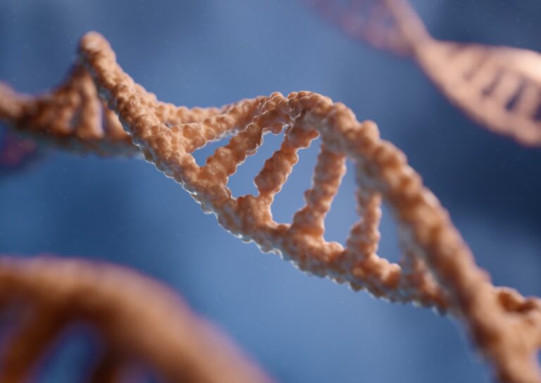
A mature human sperm cell contains 23 pairs of chromosomes. These chromosomes are then passed on to the egg during fertilization, resulting in an embryo with 46 chromosomes.
Studies of the nuclear architecture of sperm cells have been limited by their flattened nuclei. However, a combination of multicolor banding (MCB) and three-dimensional analysis of interphase cells has allowed researchers to identify chromosomes by groups.
23 chromosomes
Sperm are tiny cells that play a crucial role in reproduction. Males produce millions of these cells each day, yet it takes just one to fertilize an egg and create a new life. These cells are called spermatozoa and contain chromosomes, which are long molecules of DNA (deoxyribonucleic acid) that carry genetic information. The head of a sperm contains a cap that doctors call an acrosome and proteins that help it penetrate the outer shell of an egg. The midsection of the sperm contains mitochondria, which convert sugar into energy. The tail of the sperm, known as the flagellum, propels the sperm forward as it travels through the female reproductive tract.
A mature sperm cell, or spermatozoon, has 23 chromosomes. This is the result of a process called meiosis, which reduces the number of chromosomes in the parent cell to produce a gamete. When a sperm cell fuses with an egg cell, it results in a diploid cell with 46 chromosomes.
Sperm cells are haploid, which means they only have half the number of chromosomes found in an ordinary cell. This reduction of chromosomes occurs in specialized cells called meiocytes during spermatogenesis. After meiosis, each secondary spermatocyte has 23 chromosomes. Once the sperm and egg cells combine, they form a diploid cell with 46 chromosomes, which is known as a totipotent zygote.
The head of the sperm
In humans, sperm cells are haploid, meaning they have only one copy of each chromosome. These chromosomes determine the physical traits of the baby, including its sex (whether it is a boy or girl). The genetics of the father and mother are also passed on through the X and Y chromosomes. In order to achieve sex determination, the sperm and egg fuse in a process called fertilization.
The head of the sperm is a small, flattened cell that contains 23 chromosomes. It is shaped like an almond and is four to five micrometres long and two to three micrometres wide. The head of the sperm is made up of two parts, the sperm body and the flagellum, a slender lash-like appendage that protrudes from the head of the sperm.
The sperm nucleus is located at the center of the sperm head. It consists of a nuclear envelope and DNA, and is protected by the sperm tail. The sperm nucleus is also surrounded by the axoneme, which consists of nine doublet microtubules with two central singlets. The axoneme is important for sperm movement and is responsible for the sperm’s ability to swim. Researchers have found that mutations in the centriole protein can affect sperm head structure. If the mutation has a beneficial effect on sperm movement, it is likely to be selected.
The midsection of the sperm
Sperm cells, known as spermatozoa, play an important role in fertilizing eggs and creating new life. They are small, egg-shaped cells that contain 23 pairs of chromosomes for a total of 46 chromosomes. In addition, a sperm cell contains a DNA marker called an X or Y chromosome, which determines the sex of a future child.
Each sperm is made up of three parts – the head, neck, and tail. The head of a sperm contains the genetic material needed to fertilize an egg. It is covered by a cap-like structure known as the acrosome, which helps sperm penetrate the outer layer of an egg during fertilization. The midsection of a sperm contains tightly packed mitochondria, which provide energy for movement. It also contains a tail that propels the sperm towards an egg.
The sperm tail moves in a wave-like manner and propels the sperm towards an egg. When the sperm gets close enough to an egg, the acrosome is discarded and the X or Y chromosomes in the head of the sperm are combined with the corresponding chromosomes in the egg cell. During this process, the genes that determine a person’s physical sex are randomly selected from the 23 chromosomes in the sperm cell and the 46 chromosomes in the egg. Then, the sperm and egg combine to create a new human being.
The tail of the sperm
Sperm cells are the male gametes that enter the female reproductive tract to fertilize an egg. They form during the process of spermatogenesis, which occurs in the seminiferous tubules of the testes in mammals and other amniotes. During spermatogenesis, spermatocytes divide to produce several sperm cell precursors, including spermatogonia and spermatids. The sperm cell precursors then undergo meiosis, which reduces their number of chromosomes by half. The result is a diploid cell with 23 chromosomes, each composed of a single chromatid.
When a human sperm cell is fertilized, it becomes a zygote, a diploid egg cell. The zygote has two copies of each chromosome, resulting in 46 chromosomes. This is why it is important to determine the chromosomal constitution of a human sperm cell.
In order to study the chromosomes of a human sperm, scientists use a technique known as chromosome painting. This involves staining a sperm nucleus with various dyes. The sperm nucleus is then imaged under the microscope, and the locations of each chromosome are identified. The researchers then use a computer program to analyze the images and identify any abnormalities.
In human sperm, the chromosomes are found in a region of the nucleus called the chromocentre area. The chromocentre is located near the centrosome, and it is a site of chromatin condensation. The chromosomes are also found in a globular structure called the chromatin fibre, which is approximately 1,000 nm in diameter. The chromatin fibre is made up of a series of connected globular structures, each of which contains 3-5 Mpz DNA2.
See Also:


